Applications of Ions
Applications of ions cover quite a range of disciplines reaching from fundamental research over applied studies and industrial applications up to modern medicine. The following sections describe the fields in which DREEBIT pursues research and development: Spectroscopy, surface analysis and modification, and cancer therapy.
Spectroscopy
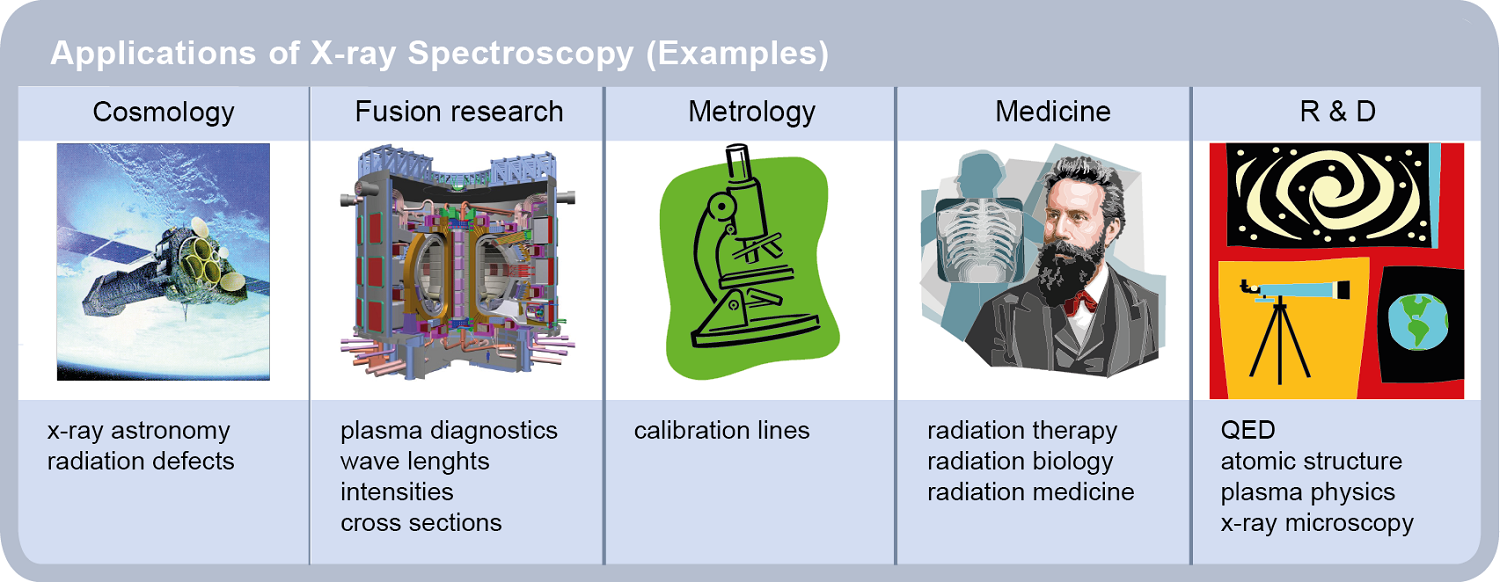
Ions created in the laboratory can be used in spectroscopical investigations for a better understanding of processes occuring in natural and artificial plasmas. Figure 1 gives an overview of the applications of spectroscopical measurements in different fields of fundamental and applied research.
Spectroscopy with DREEBIT ion sources
Since the electron energy can be sharply adjusted, expecially ion sources of the Dresden EBIT/EBIS type are excellent sources of electromagnetic radiation such as
- x-rays
- ultraviolet radiation UV/EUV
- visible light
Due to the ability of DREEBIT ion sources to ionize almost all elements up to high ion charge states spectroscopic investigations can be done for a very wide range of ion species. For an overview of the specific processes observable by spectroscopic means and the ion species which can be produced by DREEBIT ion sources please recall our article on Ion Production.
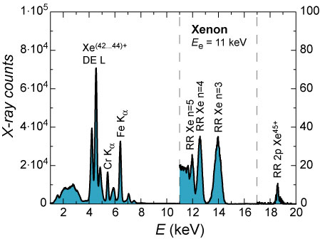
The intensity of x-rays emitted from Dresden EBIT and EBIS machines is high enough to provide energy-dispersive as well as wavelength-dispersive x-ray spectroscopy. For example, an x-ray spectrum of highly charged xenon ions is shown in Figure 2. The measurement was accomplished with a Dresden EBIT and a Si(Li) detector featuring an energy resolution of 133 eV at 5.9 keV photon energy. The left part of of the graph shows x-rays from DE processes, while the right part presents RR x-ray transitions into different subshells.
|
Figure 2 - Energy dispersive x-ray spectrum of radiation emitted by xenon ions produced in a Dresden EBIT |
|
Figure 3 - Wavelength dispersive spectrum of radiation emitted by xenon ions produced in a Dresden EBIT |
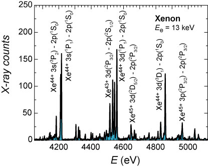
Wavelength-dispersive x-ray spectroscopy using e.g. a crystal diffraction spetrometer allows for measuring high-resolution x-ray spectra resolving manifold individual x-ray transition from different atomic states and from individual ion charge states. An example for such a measurement with xenon injected into a Dresden EBIT is given in Figure 3.
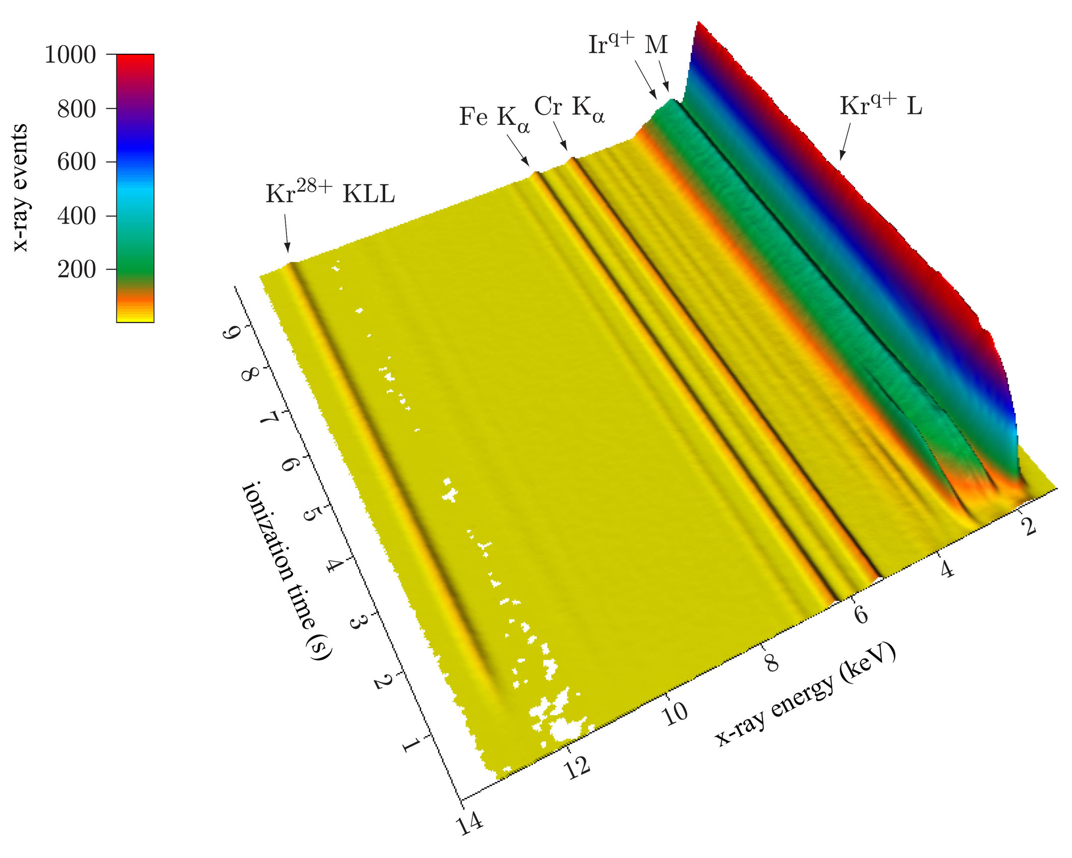
In addition to using conventional x-ray detection methods with semiconductor detectors or crystal diffraction spectrometers, DREEBIT scientists have developed a measurement system for time- and electron beam energy resolved x-ray spectroscopy, see TERX Detection System in the product category Equipment & Supplies. Some examples for measurements on x-rays emitted by krypton ions produced with a Dresden EBIT in combination with the TERX system are given in the following graphs Figure 4 and Figure 5.
|
Figure 4 - Time resolved x-ray measurement of various krypton lines including KLL-dielectronic recombination |
|
Figure 5 - Electron beam energy resolved x-ray measurement of krypton KLL-dielectronic recombination lines |

Surface Analysis and Modification
Ion beams are well-approved tools for the analysis and modification of surfaces in research as well as industry. Probably, the most well-known example is the application of ion beam implantation for the production of semiconductor devices. In this field, ion beams are mostly scanned over surfaces or widened to cover large wafer areas. New developments in applied research also tend towards focussing ion beams to very small spots with the ultimate goal of the direct production of nano-scale devices and circuits. DREEBIT ion sources and ion beam facilities have been used for several applications in this field as described in the following.
DREEBIT Ion Sources for Nanoscale Surface Modification and Analysis
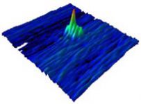
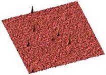
Since 2003, Dresden EBIT/EBIS machines have been used by several research groups for the production and studies of nanostructures. Figure 6 and Figure 7 show two examples for nanodots produced by irradiation of surfaces with highly charged ions from Dresden EBIT ion sources.
|
Figure 6 - HOPG 20 nm x 20 nm irradiated with Xe20+ at 4 keV (by courtesy of Dr. Facsko, Helmholtz-Zentrum Dresden-Rossendorf) |
Figure 7 - MICA 250 nm x 250 nm irradiated with Ar16+ at 8 keV (by courtesy of Prof. Schleberger, University of Duisburg-Essen) |
The interest of these fundamental studies is directed towards understanding how exactly the surfaces are modified by the kinetic and potential energy of the incident ions to be able to create nanostructures with specific properties which can be used in future electronic devices. More information and links to papers about these studies can be found in our section about References, see "Dresden EBIT and EBIS-A at HZDR" and "Dresden EBIS-A and Dresden EBIT at University of Duisburg-Essen".
Focused Ion Beam (FIB) Machines and Time-Of-Flight Secondary Ion Mass Spectrometry (TOF-SIMS)
Next to understanding the basics of ion-surface interactions in detail other studies concern the localization of the ions impacting the surfaces. So-called Focused Ion Beam (FIB) machines include lenses which effect the focusing of the ion beam down to a minimal spot size in the nanometer or sub-nanometer range which is determined by the characteristics of the focusing system as well as by the properties of the employed ion source. FIB machines equipped with Liquid Metal Ion Sources (LMIS) are already in use for industrial applications, e.g. for the preparation of lithography masks in semiconductor factories.
LMIS are sources of very high brilliance but only produce low charged ions. One of the on-going R&D activities at DREEBIT laboratories is the operation and enhancement of experimental FIB machines working with specifically designed Electron Beam Ion Sources (EBIS) which are able to produce Highly Charged Ions (HCI). With these new HCI-FIB devices, the effect of the stored potential energy in HCI can be exploited for the strongly locallized irradiation of targets. This is of interest for the production of quantum dots, a nowadays largely discussed research topic. However, the spot size of HCI-FIB machines currently lies in the range of several 10 micrometers which is still to be improved. Figure 8 shows the operation principle of the setup at the DREEBIT laboratories and the "D" from DREEBIT written with an Ar8+ beam. The image of the written structure was generated at the same setup by scanning the surface again with an ion beam and reading out the information from a Secondary Electron Multiplyer (SEM).
|
Figure 8 - Operation principle of the DREEBIT HCI-FIB and an SEM image of a structure written by an argon ion beam. |

Next to the modification of surfaces, the HCI-FIB machine can thus be used for position-sensitive analysis of targets. In the SEM imaging mode, a resolution in the micrometer and sub-micrometer range has already been achieved. Furthermore, the DREEBIT Time-Of-Flight Secondary Ion Mass Spectroscopy (TOF–SIMS) setup was added to the HCI-FIB machine for mapping surfaces including information about the chemical composition of the material. TOF-SIMS uses the incident pulsed ion beam to generate secondary ions at a small spot on the sample surface which are extracted and accelerated in a well defined potential. The accelerated ions are detected at the end of a certain drift space and identified according to their time-of-flight which is correlated to a certain mass-to-charge ratio.
For FIB and TOF-SIMS applications the use of Dresden EBIS for the production of HCI provides a number of outstanding features such as a large spectrum of available primary ion species. Operators have a wide choice to optimize the charge and mass of the projectiles for a certain material to achieve the highest possible rates of secondary particles for a very sensitive surface analysis. If you are interested in working with the HCI-FIB / TOF-SIMS, DREEBIT offers beamtime for guest scientists as well as engineers working in research and development. For more information about our user facilities please visit our Service section or Contact us directly.
IMALION - a DREEBIT Facility for Ion Irradiation of Large Surfaces
Imalion stands for the IMplantation of ALuminum IONs since the facility was originally developed for aluminum implantation into wafers. The ECR ion source implemented in the machine is, however, not restricted to only produce aluminum ions but covers a large spectrum of low charged ions with high intensities in the milliampere range. For more information on DREEBIT ECRIS please see our product section Ion Sources.
|
Figure 9 - Overview of the IMALION facility |
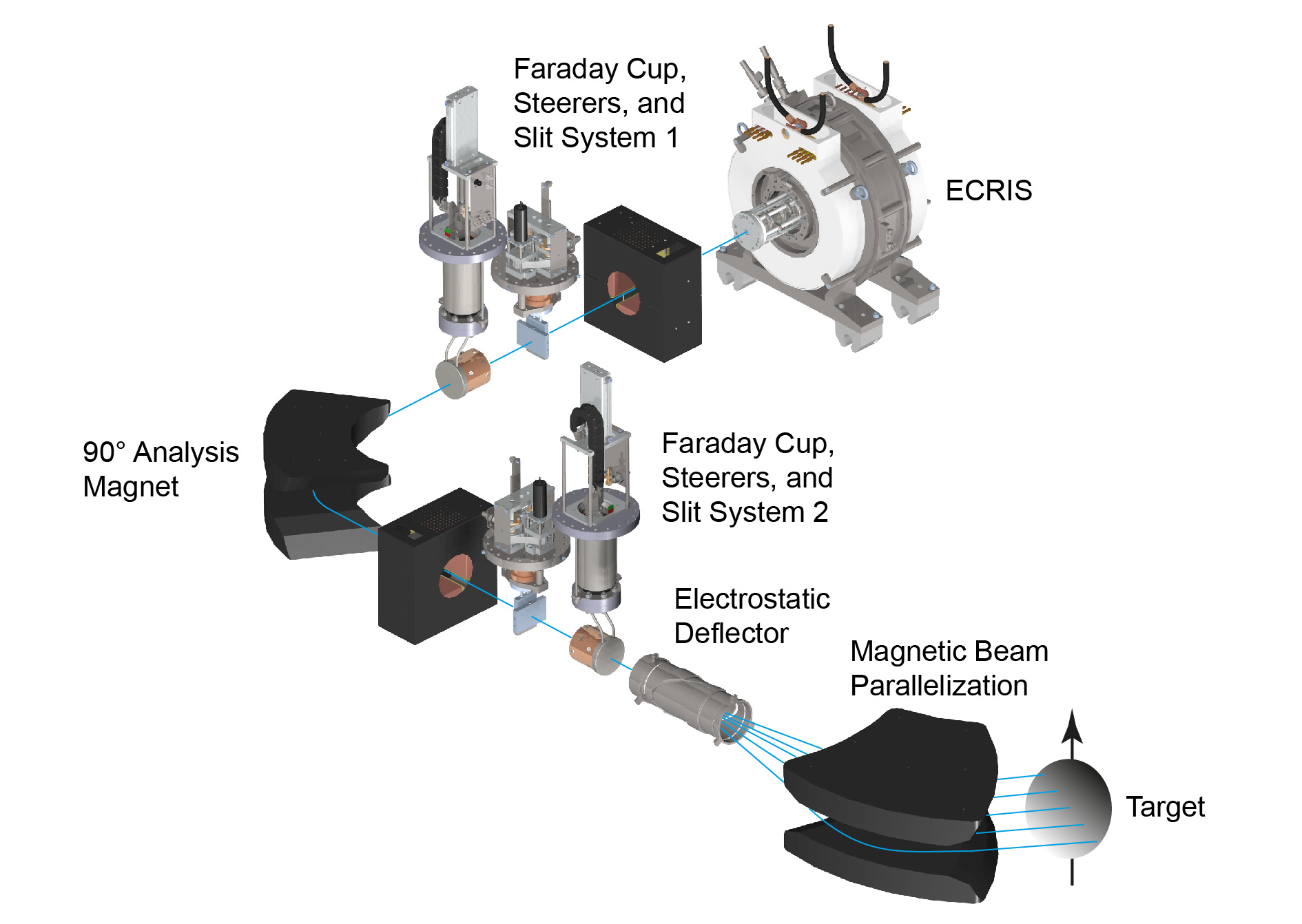
All components of the IMALION beamline, shown in Figure 9, were developed by DREEBIT scientists and engineers. The basic beamline elements include beam optics such as magnetic steeres for correction of the beam direction as well as beam diagnostics like the 90° bending magnet for charge-to-mass ratio separation and high-current faraday cups for the detection of the electric beam signal. To realize the irradiation of large surfaces the Wide Area Irradiation Optics WAIO were developed consisting of an electrostatic deflector to scan the beam in one direction and simultaneously re-focus the beam for injection into a parallelization magnet which ensures a perpendicular angle of incidence onto the target.
As in the case of DREEBIT's HCI-FIB /TOF-SIMS setup, beamtime can be booked at the IMALION facility as well. If you are interested please visit the section Service or Contact us for more information.
Medical Particle Therapy
Cancer is the second most common cause of death worldwide and about 33% of all inhabitants of the European Union will be confronted with some kind of cancer in their lives. This rises the demand for more treatment facilities and for higher quality treatments to reduce undesirable side effects. Currently about 45% of cancer cases can be treated, mainly by surgery and / or by radiation therapy.
Aside from surgery, radiotherapy is the main treatment of cancer nowadays. Within the radiotherapy, the radiation with hadrons (mainly protons and carbon ions) is the most promising treatment technique. Up to now (2012) about 71.000 patients worldwide were treated by particle therapy at 32 institutions (Europe, USA, Japan, China, South Africa) and the number of treated patients increases constantly. For a list of the particle therapy centers in operation and under construction please visit the website of the Particle Therapy Co-Operative Group, PTCOG.
Cancer Treatment with Ions Beams
The classical radiation technique uses x-ray photons to destroy tumors. Unfortunately, the photons interact strongly with the healthy tissue along their way to the tumor. The most dose is applied into the first centimetres of the patients tissue. For inner tumors the ambient healthy tissue is more damaged than the cancer itself. In order to reduce the damage of the healthy tissue it is standard to rotate the x-ray emitter around the patient with the tumor in the focal point of the emitter.
|
Figure 10 - Applied dose versus depth for various tumor irradiation methods |

In contrast to the classical x-ray radiation the treatment with ions - in particular with proton and carbon ions - have significant advantages. The energy deposition within the tissue obey to the Bragg interaction. I.e. the maximum of the energy is deposited at the end of the particle trajectories, hence in the tumor cells. The ion range can be adjusted easily be varying the kinetic energy of the particles. Due to the Bragg interaction the healthy tissue is spared (important for tumors in the near of risky organs like the medulla).
Another important property of ions as opposed to photons is their electrical charge. Charged particles can be focused and deflected by electric and magnetic fields, providing the ability to scan with the ion beam over the tumor very precisely. Due to the variation of the kinetic energy of the ions it is possible to vary the penetration depth. Hence the tumor can be scanned voxel by voxel (a voxel is a three dimensional volume element).
Beside the electrical properties the relative biological efficiency (RBE) of the ions is higher by a factor of about three. This means that the same dose of carbon ions effects the tumor tissue by a factor of 3 more compared with classical x-rays. The higher RBE allows a lower dose by the same effect on the tumor with less damage of the healthy tissue.
For the treatment of tumors deeply located in the tissue total kinetic energies of up to 250 MeV for protons and up to 4.8 GeV for carbon ions are required. Hence the ions have to be accelerated by high-performance particle accelerators. The state-of-the-art production of ions before acceleration is performed using Electron Cyclotron Resonance Ion Sources (ECRIS). However, the use of Electron Beam Ion Sources (EBIS) is under discussion as an improvement for ion therapy centers of the future.
Application of DREEBIT Ion Sources in Particle Therapy
DREEBIT follows two approaches of introducing new ion sources in medical cancer therapy with remarkable advantages compared to classical ECRIS technology. The compact and easy-to-use Dresden EBIS-A, a room-temperature electron beam ion source of small dimensions and few infrastructural requirements, provides enough performance to be used in up to 70% of the treatment scope. It is a powerful addition to ECR ion source based facilities with low operation costs and high operation stability.
On the other hand, the Dresden EBIS-SC is a compact superconducting EBIS which is based on the most modern principles of refrigeration as well as electron beam technologies. On-going studies investigate the advantages of completely replacing ECR ion sources by this kind of electron beam ion source. For more information about the Dresden EBIS-SC used in particle therapy please see our recent publications, e.g. by Ritter et al. as part of the proceedings of the IPAC conference held in 2014.
After test runs carried out at DREEBIT laboratories, the Dresden EBIS-SC was transferred to the Heidelberg Ion-Beam Therapy Center HIT in 2013 to evaluate the feasability of using the superconducting EBIS for medical particle therapy directly on-site. An image of the EBIS-SC installed at the HIT test bench is given in Figure 11. Among others, the most promising result of the studies conducted at HIT was that the emittance of the Dresden EBIS-SC is lower by a factor of nine compared to a traditionally used ECRIS which improves the transmission through the following accelerator structures meaning that less ions are lost during transportation to the patient. Therefore, this kind of therapy can be made more efficient and affordable by using the EBIS instead of an ECRIS.
|
Figure 11 - The Dresden EBIS-SC installed at the HIT ion source test bench |
Furthermore, many medical irradiation facilities are completely occupied by particle therapy at daytime on workdays but during nights and weekends the accelerator can be used for non-clinical research, e.g. for nuclear physics, radiation biology, materials research and others. For these activities many different ion species are desired and should be available from the installed ion source with the same q/A ratio. In this case, EBIS can ionize heavier elements with the same q/A ratio cost-efficiently and with appropriate ion beam currents. In comparison to classical ECR ion sources this is another significant advantage with convincing scientific and economic aspects.